
Fixed Partial Dentures (FPDs) supported by both implants and natural teeth
In cases of partial edentulousness where implant supported bridges, ISBs, are planned in the lateral segments of the jaws, the amount of available bone often limits treatment possibilities. In the maxilla, recesses in the sinus maxillaris fill out the toothless alveolar process, and in the mandible, resorption reduces the distance from the alveolar ridge to the n. alveolaris inferior. This makes the placement of fixtures, in sufficient number and of sufficient length to support an ISB, more difficult.
Methods have been developed for transplanting bone to the sinus maxillaris and transposing n. alveolaris inferior. These methods are, however, combined with a more demanding, lengthy treatment and an increased risk for complications. In cases suitable for this procedure, e.g. missing teeth behind the canine, a simpler method can be appliedfor replacing the premolars.
In the mandible, a long, oblique fixture can be placed between the foramen mentale and the apex of the canine, opening in the alveolar process in the region of the second premolar.
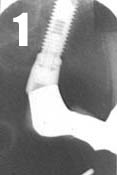     |
In the case presented, with treatment of
a maxilla, a fixture is placed in front of the sinusí frontal border. By angling the
fixture posteriorly, it is possible to fit a long fixture which opens in the region of the
second premolar (Fig. 1). After 6 months healing, the abutment connection can be made. In
this case, a 32° angulated abutment is used.
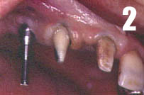
After preparing and laying open the supporting teeth, in this case nos. 13 and 11, the
abutment is provided with an impression coping (Fig. 2). An impression is taken with an
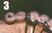
elastomer. The bridge is made of porcelain fused to metal with an intracoronal attachment,
in this case McCollum, and with a palatinally placed horizontal locking screw which
prevent vertical gliding (Fig. 3). The construction allows the implant-supported portion
of the bridge to be taken off when necessary, e.g. when tightening the abutment screw. The
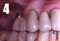
bridge is cemented in the usual manner on the supporting teeth with simultaneous
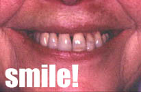
tightening of the gold screw in the implant-supported section. The bridge is cemented
(Fig. 4), smile! (Fig. 5).
The requirement which ought to be fulfilled is that the bridge be supported by a fixture
placed at a maximum of one tooth width from a supporting tooth. The toothís mobility
ought not exceed the physiological mobility. With the correct indications, this treatment
is both time-saving and cost-reducing.
References
1. Rangert B, Gunne J, Sullivan D. Mechanical aspects of Brĺnemark implants connected to
a natural tooth: An in vitro study. J of Maxillofacial Impl 1991; 6:177-186.
2. Gunne J, Ĺstrand P, Ahlén K, Borg K, Olsson M. Implants in partially edentulous
patients. A longitudinal study of bridges supported by both implants and natural teeth.
Clin Oral Impl Res 1992; 3:49-56.
Responsible Operators
Implant surgeon Nils Ravald DDS, PhD Simon Dahlgren, DDS, Specialist in Prosthodontics
Specialist Center for Oral Rehabilitation, Linköping, Sweden