
by Leif Kullman
Computed tomagraphy
Principle
CT, or computed tomography, is nowadays a routine procedure in medical imaging that produces images of the internal tissues of the human body. The main feature of the CT scan is the bore (figure 1), a wide opening through which the patient moves. The patient is lying on the investigation table of the scanner. As the patient passes through the bore the x-ray tube rotates in a helical, or spiral motion round about the patient 360 degrees and takes a "picture" or "slice". After many such "slices" the computer has enough information to combine various segments of the pictures and create views of the internal organs of the body. Computererized pictures are received that reflect the energy absorbed by the body tissues in a usual radiographic way. These views are projected onto a monitor and are recorded on film.
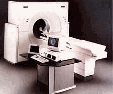
Figure 1.
The produced images are cross-sectional and their pattern (figure 2) can be compared with slices of a loaf of bread. The examination time depends upon area of the body being viewed and can be from 15-20 minutes to 2 or 3 hours. Patients who cannot lie still for that period of time require sedation to help them being motionless.
By taking a series of such images, a CT can create a multidimensional (3-D or three dimesional) view of the body. Now the whole loaf of bread can be seen! The quality of this multidimensional view enhances the radiologist's ability to accurately diagnose different pathological conditions.
3-D image reconstruction also permits three-dimensional processing and display of CT image data for surgical planning, such as orthopaedic prosthesis, or facial-cranial reconstruction. Displays include patient volume in surface or volumetric three-dimensional rendering.
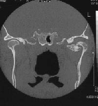
Figure 2: A frontal or coronal slice through the temporomandibular joint area of the skull. An osteochondroma can be seen in the left medial side of caput mandible.
To improve the ability to make correct diagnosis, contrast media are very often used. These media contain iodine or barium which x-rays cannot penetrate. By ingesting or injecting this media into the body the radiologist can better visualize structures that may not otherwise be seen. Contraindications to many contrast media include pregnancy, nursing, iodine-related allergies or kidney disease.
Usually used CT scans of the different body organs are:
1. Head scanning: looks at the skull bones for possible fracture; Brain for possible stroke, bleeding, or abcesses. Evaluating the jaws before implant installation.
2. Back scanning: To look at the anatomy of the spinal column such as spinal stenosis (tightness around the spine), possible fracture, or nerve impingement .
3. Chest scanning: To look at the lungs usually evaluating an abnormality first seen on a chest x-ray.
4. Abdomen and Pelvis scanning: To look at the different organs in the upper and lower abdomen.
Next month I will continue to show radiographic images describing some cases of different pathology.
Leif Kullman